As in every part of the world today we celebrate the fight that many women had won agains breast cancer. This helps to rise attention, and support related to the awareness, early detection, treatment, and palliative care.
According to the OMS, breast cancer generates 1.38 million new cases each year and 458000 women dead due to that illness, the more registered in development countries due to the late detection lack of sensibility and the difficulty to access to health services.
Did you know that an early detection of cancer save lives and reduce the cost of the treatment?
Prevent is living
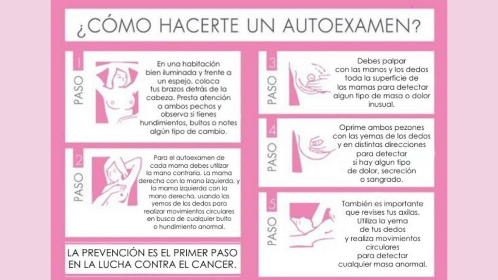
Knowing well your breast is a vital importance and there’s nothing better for that than a self expiration every month, because if any change is found earlier it could save your life.
The self exploration is a self exploring method based on the checking of the breast done by the woman herself.

Picture by Contra piel
The best time to do a self exploration is once the period is over. It’s a commitment for health staff or nurseries of the health centre you’re going to, to teach you how to explore yourself and provide you information about symptoms and any breast cancer signs, touch yourself each month learning to know it’s consistency, texture, shape and develop a more sensitive sensibility on our hands which would help us to identify any change.
Clinic breast exploration
When you go to see a doctor he/she would offer you the “clinic breast exploration”, which consist on a two phase thing one is the observation and then the touching.
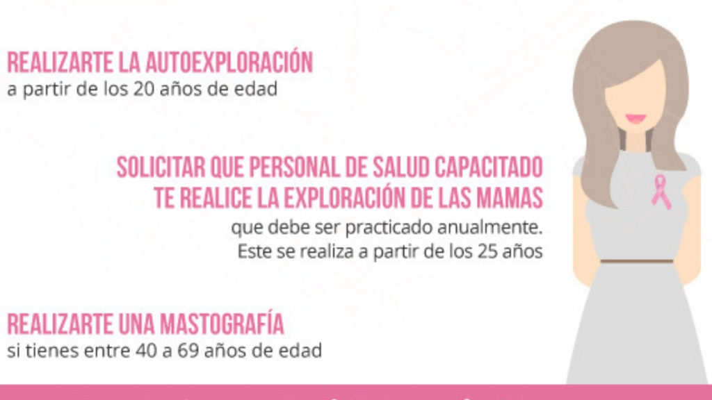
If the exploration is a regular one we recommend you to repeat it the next year. If you find anything wrong then you will ve sent to your family doctor for a new checking. This procedure should be started at the age of 25.

Picture by IMSS
Non invasive diagnostic proofs
Its a breast radiography. Realized with an X-ray machine specially designed for breast, and with a really low dosis of radiation it’s capable to find any problem, mainly breast cancer.
The mammograph is placed in the breast area, and the breast is slightly pressed during the test, but this is not painful at all. The information that it realizes due to neo formations, micro calcification, and distortions in the breast tissue could lead us to a definitive diagnostic.
Breast ultrasound is something that actually complements the first procedure on many occasions, intramammary structures such as cysts can be described in greater detail.
Ultrasound is also indicated for young or high density breasts. The doctor slides gently on the breast a probe that emits ultrasonic waves. When crossing the tissues, these waves bounce off them, producing ultrasonic echoes that are represented on the screen of the ultrasound and can be photographed.
It is a magnetic resonance of the breast. It is used as a complementary study to the previous tests and for high risk patients.
Risk factor’s
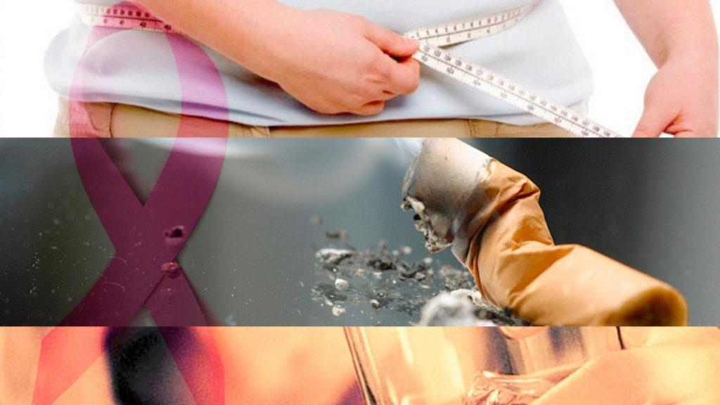
Some studies have shown that the risk of having breast cancer is due to a combination of factors. The main factors that influence a person’s risk include being a woman and getting older, others are the following:
- High fat diets
- Obesity
- Consumption of alcohol
- Hormone replacement therapy for more than 10 years

Picture by Vallarta Opina
If you have risk factors for breast cancer, consult a doctor so that you are aware of the ways in which you can reduce the risk of developing this disease.
Download TinkerLink for free! And find a trustful doctor trained in the detection and prevention of breast cancer!
[vc_column][ultimate_info_banner banner_desc=»You might be interested in: Tips for a sustainable lifestyle» button_text=»Read Now» button_link=»url:%2Ftips-para-llevar-una-vida-sustentable%2F|||» info_effect=»fadeIn» button_color=»#9c27b0″ button_border_width=»1″][/vc_column]


